Synapse

Google Search of SETI Net
Synapses
The connection between two neurons is called a synapse. Since each neuron can connect to thousands of other neurons, each neuron can have many thousands of synaptic connections.
Synapses are physically at the tip of the Dendrite. This is the location where the pulse of chemicals, the action potential, is converted into the release of neurotransmitters. The Synapse membrane is less than 50 nanometers away and contains specialized receptors. The neurotransmitter rapidly (in microseconds) diffuses across the synaptic cleft and binds to specific receptors .
The type of neurotransmitter released from the terminal and the specific receptors present on the synapse are critical in determining the quality and intensity of information transmitted by neurons.
Excited and Depressed Dendrites
There are two radio buttons labeled EPSP and ISPS. If IPSP is set, the synapse acts as an Inhibitory Postsynaptic Potential input to the dendrite cell.[kandel page 275 *] . If EPSP is set, the synapse is connected to an EPSP synapse. These connections are visualized in the following image.
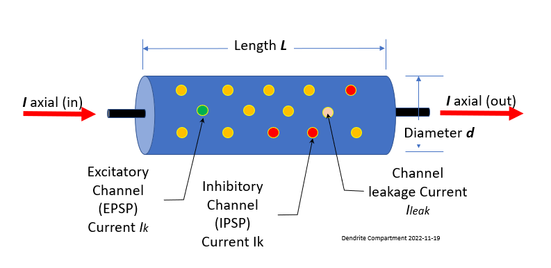
Connections from other neurons Axons may be attached to any one of thousands of places along the dendritic tree. The drawing of a compartment of dendrite (above) shows two types of ion channel connections:
- EPSP, Green - If the connected ion channel stimulates output, it is called Excitatory Post-Synaptic Potential
- IPSP, Red - If the connection channel depresses the downstream soma from firing it is called an Inhibitory Post-Synaptic Potential .
Each of these are connected through synapse. Leak channels shown in Orange are never connected.
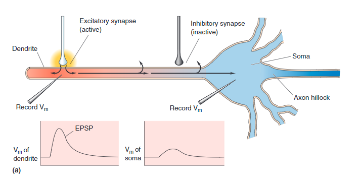
EPSP Activated
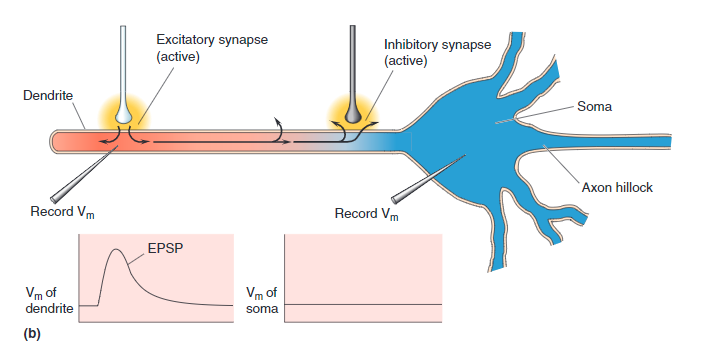
IPSP Activated
A single synapse contributes to the postsynaptic neuron's membrane potential, although the magnitude of this contribution depends on several factors:
- Synaptic Type (Excitatory or Inhibitory)
- Synaptic Current
- Membrane time and Space Constants
- Synapse location on the Dendritic Tree
- Temporal Factors ( decay and summation)
It works out that a single synaptic event when connected to an IPSP channel moves the Dendrite membrane level about 3.24 mV. When connected to an EPSP channel it moves -3.24 mV.
The Neuron Lab simulator does not simulate to the level of the synapse neurotransmitters. In this simulator, the synapses' role is in support of the phenomenon of Spike Timing Delay Plasticity (STDP), which is described in the section on STDP.
The Simulator
If you right-click on a Synapse icon, you are presented with the internal workings of the Synapse. This Synapse is receiving 1 pico Amp at its input and producing 3.18 millivolts at its output to the following dendrite section.
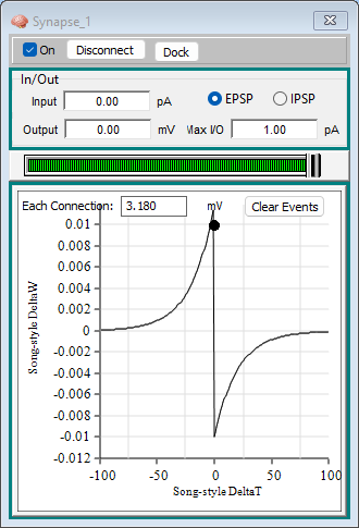
The bottom curve is showing the STDP results for this synapse. More on STDP in the following section ( STDP ). You can pull the synapse up from the bottom to ignore this.
This 3.18 mV output is the maximum output for a single synaptic connection. If you wanted to simulate raising the Dendrites membrane to be enough to trigger an output from a following Soma (depolarizing from -60 mV to -40 mV), you would have to connect eight different synapses. To facilitate a more reasonable connection setup, the simulator allows higher input currents.
For example:
Install the neuron simulator, if you have not already, and create the setup this simple connection.
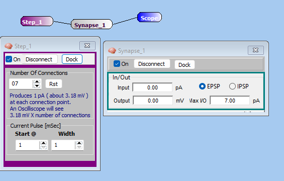
The scope will show a very small input caused by 1 pA presented simultaneously to 7 different equivalent synapses.
To be biologically plausible, only a single Synapse could connect to a single Dendrite receptor. The simulator relaxes these rules by allowing any number of connections.